Translate this page into:
Cystoid Macular Edema Secondary to Paclitaxel Therapy

Corresponding Author: Anibal Martin Folgar, Universidad Maimonides, Oftalmologia, Hidalgo 775, Caba, Buenos Aires-1407, Argentina. Email: anibalmartinfolgar@gmail.com
-
Received: ,
Accepted: ,
How to cite this article: Folgar AM, Zarate JO. Cystoid macular edema secondary to paclitaxel therapy. Lat Am J Ophthalmol 2019;2(1):1-4.
Abstract
We present a 57-year-old referred reduced visual acuity who was in treatment with paclitaxel for developing metastatic breast adenocarcinoma. Ophthalmoscopic examination, optical coherence tomography, and autofluorescence show the cystoid macular edema, but fluorescein angiography is normal, without leakage of dye in the late times. The patient responds well 8 weeks after stopping antineoplastic. Paclitaxel can cause cystoid macular edema and lifting a recovery both anatomical and functional of the macula.
Keywords
Cystoid macular edema
Fluores-cein angiography
Paclitaxel
INTRODUCTION
The cystoid macular edema is a sign that is revealed by capillary leakage in the late times of the angiogram. When this is not evident, it is a rare condition. There are reports that show this clinical feature mainly in nicotinic acid maculopathy.[1-5] Recently, there have been cases reported as an adverse effect associated with a family of taxane antineoplastic agents, such as paclitaxel docetaxel.[6-13]
CASE REPORT
Female patient, 57 years old. Which refers by gradual decrease visual acuity in both eyes of 1 month of evolution. She presents as a pathological antecedent, locally advanced tumor of the left breast (Biopsy on March 20, 2010) T4a N2 IIIB M0 Slightly differentiated infiltrative carcinoma ER (positive estrogen receptors) in 40% of the cells; RP (progesterone receptors): positive in 10% of the cells; Immunomark +++ for HER 2/neu oncogene (c-erb-B2): (Score 3).
Chemotherapy begins on May 17, 2010 to April 4, 2010 with (4 cycles), 1 cycle every 21 days for 4 cycles off AC (doxorubicin/cyclophosphamide) 60 mg/m2/ 600mg/m2. Subsequently, between August 26, 2010 and April 29, 2011 , she received 11.6 cycles of Trastuzumab (Herceptin, Genentech Inc.), 4mg/kg of attack dose and 2mg/kg of maintenance dose plus Paclitaxel 90mg/m2 weekly (Taxol, Bristol-Myers).
In the initial eye, examination showed a best-corrected visual acuity (measured with early treatment diabetic retinopathy poster) which was 20/40 in both the eyes. The biomicroscopy of the anterior segment examination was normal, intraocular pressure was 14 mm/Hg in both the eyes, and the eye fundus examination revealed cystoid macular edema.
The digital fluorescein angiography performed with a TRC-50DX and IMAGEnet capture software (Topcon Medical Systems, Inc.) showed choroidal filling and retinal vascular on both normal late times showed no dye leakage.
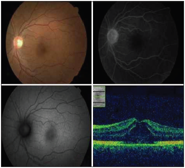
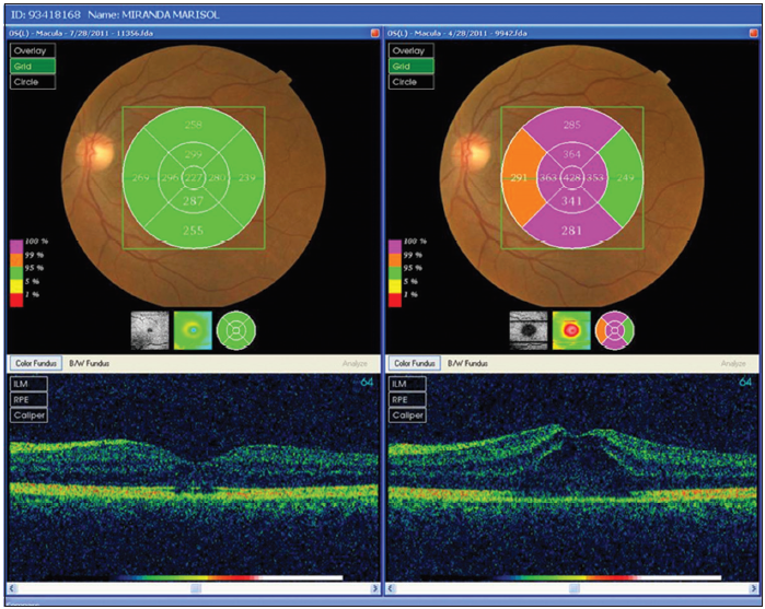
The optical coherence tomography (OCT) performed with spectral domain 3D OCT-2000 (Topcon Medical Systems, Inc.) showed cystoid macular edema with a central macular thickness of 459 um for the right eye and 428 um for the left eye. The fundus autofluorescence performed with a TRC-50DX and Spaide FAF filters (Topcon Medical Systems, Inc.) revealed hyper autofluorescence on multispot pattern in both the eyes [Figures 1 and 2]. The subject was treated with a multidisciplinary oncology service, and discontinuation of paclitaxel was indicated.

- Female patient, 57years old. Who complains of gradual decrease in visual acuity in both eyes 1month of evolution. In this picture we can see a collage of the right eye, optical coherence tomography showed cystoid macular edema the fundus autofluorescence revealed hyper auto fluorescence on multi-spot pattern in both eyes and does not exhibit any signs of fluorescein leakage in the angiography fluorescein

- Female patient, 57years old. Who complains of gradual decrease in visual acuity in both eyes 1month of evolution. In this picture we can see a collage of the left eye, optical coherence tomography showed cystoid macular edema the fundus autofluorescence revealed hyper auto fluorescence on multi-spot pattern in both eyes and does not exhibit any signs of fluorescein leakage in the angiography fluorescein
The patient evolved favorably, and within 8 weeks, best-corrected visual acuity had improved to 20/20 in both the eyes. In biomicroscopic examination of the fundus, macular edema is not evident. The optical coherence tomography It showed a thickness normal foveal center (od: 223 um; oi: 227 um) with a focal alteration in the line of union between the internal and external segments of the photoreceptors [Figure 3].

- Female patient, 57years old. Who complains of gradual decrease in visual acuity in both eyes 1month of evolution. In this picture we can see after 8weeks in the optical coherence tomography showed a center normal thickness and a focal abnormality in the connection line between the inner and outer segments of photoreceptors
DISCUSSION
Taxanes are terpenes produced by plants of the genus Taxus as Tejo, hence its generic name. They were first identified in natural sources, but some of them have been artificially synthesized. Paclitaxel and docetaxel are taxanes. The taxanes are antimicrotubule agents promotes the assembly of microtubules from tubulin dimers and stabilizes microtubules by preventing depolymerization.
This stability results in the inhibition of the normal dynamic reorganization of the microtubule network that is essential for vital interphase and mitotic cellular functions. Paclitaxel induces abnormal arrays or “bundles” of microtubules throughout the cell cycle and multiple asters of microtubules during mitosis, and this results in the inhibition. Stability of the usual dynamic reorganization of the microtubule network is essential for that vital interphase and mitotic cellular functions.
Paclitaxel induces abnormal arrays or “bundles” of microtubules throughout the cell cycle and multiple asters of microtubules during mitosis, which also has antiangiogenic effect and increases apoptosis (oncogene bcl-2).
It is used as an antineoplastic agent in solid tumors and non-solid, and their main adverse effects are myelosuppression, dermatitis, fatigue, alopecia, and mucositis.
Both docetaxel and paclitaxel[6-9,12-19] have been associated with cystoid macular edema without fluorescein leakage in the late stages of the angiogram. The pathophysiology of this type of cystoid macular edema without capillary leak in the late times of fluorescein angiography is unclear but could relate to various pathophysiological situations, such as the interstitial fluid pressure (IFP) which is important to maintain a constant volume of interstitial fluid. The reduction in IFP appears to be involved a dynamic-mediated 1-integrins and interactions between cells and fibers of connective tissue extracellular matrix (ECM). The 1-integrins are adhesion receptors responsible for binding of connective tissue cells: The ECM and the cytoskeleton.
Disruption of actin filaments leads to a decrease in IFP and edema formation, suggesting a role for actin filaments. Brønstad et al.,[19] in a study, on an animal model to investigate thoroughly the role of cytoskeleton in control of IFP studied the effect of fixation of microtubules with paclitaxel and docetaxel. Moreover, the formation of edema determined that the fixing of microtubules with taxanes increases the ability of these efforts.
This would make the contractility of the actin filaments which is less efficient and in turn causes a voltage drop in the transmission networks of the ECM, thus allowing the matrix to swell, resulting in a reduction in PFI with consequent edema.
Another effect that was observed was the capillary membrane destabilization allowing an increase in the 3 output of albumin.[19] Joshi et al.[9] and Nakao et al.[17] proposed pathophysiological mechanism of the direct toxicity of Muller cells with the consequent accumulation of intracellular fluid and a very slow rate of leakage to the extracellular space.
Another mechanism would involve a partial compromise of the blood–retinal barrier. This would make small molecules and proteins that can diffuse more readily than larger molecules of fluorescein. This would be disseminated to a much slower rate of fluorescein molecules, resulting in no apparent diffusion of dye in the angiogram late times.[11] To date, been reported in the literature. 10cases of cystoid macular edema secondary to the administration of paclitaxel[6-9,12-19] in one case are man with metastatic gastric cancer and nine cases were women who had with advanced metastatic breast adenocarcinoma and one metastatic skin melanoma [Table 1]. The cystoid macular edema resolved on discontinuation of paclitaxel in 8cases. In one of the cases[7], acetozolamide was administered orally for 6 weeks plus suspension of Paclitaxel, which also resulted in the disappearance of symptoms.
| Authors | Sex | Age | Type of cancer | Treatment | OCT | RFG | AF |
|---|---|---|---|---|---|---|---|
| Joshi et al. 2007 | Female | 63 | Breast + MMTs | Discontinue | Yes | Yes | No |
| Smith et al. 2008 | Female | 56 | Breast + MMTs | Discontinue | Yes | Yes | No |
| Murphy et al. 2010 | Female | 65 | Breast + MMTs | Discontinue | Yes | Yes | No |
| Ito et al. 2010 | Female | 62 | Breast + MMTs | Discontinue | Yes | Yes | No |
| Baskin et al. 2011 | Female | 40 | Breast + MMTs | Discontinue | Yes | Yes | No |
| Koo et al. 2012 | Female | 54 | Gastric + MMTs | Discontinue + Methazolamide | Yes | Yes | No |
| Meyer et al. 2012 | Female | 62 | Skin melanoma + MMTs | Discontinue + Acetazolamide | Yes | Yes | No |
| Modi et al. 2013 | Female | 61 | Breast + MMTs | Discontinue | Yes | Yes | No |
| Da Costa et al. 2015 | Female | 63 | Breast + MMTs | Discontinue | Yes | Yes | Yes |
| Nakao et al. 2016 | Male | 66 | Gastric + MMTs | Discontinue | Yes | Yes | No |
| Bassi et al. 2017 | Female | 49 | Ovarian + MMTs | Discontinue | Yes | Yes | No |
We can say that the dissociation between OCT and fluorescein angiography gives us a significant fact in the differential diagnosis of this type of macular edema. This is the first reported case in which was included as a supplementary study in macular auto fluorescence which turned out to be positive; this is a non-invasive test with coherence tomography providing us with an important diagnostic approach.
Note that, this type of adverse events in patients treated with taxanes is unusual. Its pathophysiology remains unclear, and further studies are needed to back up their hypothesis.
References
- Cystoid macular edema secondary to nab-paclitaxel therapy. J Clin Oncol. 2010;28:e684-7.
- [CrossRef] [PubMed] [Google Scholar]
- A case of cystic maculopathy during paclitaxel therapy. Nippon Ganka Gakkai Zasshi. 2010;114:23-7.
- [CrossRef] [Google Scholar]
- Cystoid macular edema secondary to albumin-bound paclitaxel therapy. Arch Ophthalmol. 2008;126:1605-6.
- [CrossRef] [PubMed] [Google Scholar]
- Cystic maculopathy with normal capillary permeability secondary to docetaxel. Optom Vis Sci. 2003;80:277-9.
- [CrossRef] [Google Scholar]
- Cystoid macular edema with docetaxel chemotherapy and the fluid retention syndrome. Semin Ophthalmol. 2007;22:151-3.
- [CrossRef] [PubMed] [Google Scholar]
- Macular edema associated with nicotinic acid (niacin) JAMA. 1998;279:1702.
- [CrossRef] [PubMed] [Google Scholar]
- Adverse ocular effects associated with niacin therapy. Br J Ophthalmol. 1995;79:54-6.
- [CrossRef] [PubMed] [Google Scholar]
- Abraxane-induced cystoid macular edema refractory to concomitant intravenous bevacizumab. Can J Ophthalmol. 2011;46:200-1.
- [CrossRef] [PubMed] [Google Scholar]
- Effects of the taxanes paclitaxel and docetaxel on edema formation and interstitial fluid pressure. Am J Physiol Heart Circ Physiol. 2004;287:H963-8.
- [CrossRef] [PubMed] [Google Scholar]
- Regression of paclitaxel-induced maculopathy with oral acetazolamide. Graefes Arch Clin Exp Ophthalmol. 2012;250:463-4.
- [CrossRef] [PubMed] [Google Scholar]
- Acase of paclitaxel-induced maculopathy treated with methazolamide. Korean J Ophthalmol. 2012;26:394-7.
- [CrossRef] [PubMed] [Google Scholar]
- Non-leaking cystoid maculopathy secondary to systemic paclitaxel. Ophthalmic Surg Lasers Imaging Retina. 2013;44:183-6.
- [CrossRef] [PubMed] [Google Scholar]
- Multimodal imaging in paclitaxel-induced macular edema: The Microtubule Disfunction. Cutan Ocul Toxicol. 2015;34:347-9.
- [CrossRef] [PubMed] [Google Scholar]
- Possibility of müller cell dysfunction as the pathogenesis of paclitaxel maculopathy. Ophthalmic Surg Lasers Imaging Retina. 2016;47:81-4.
- [CrossRef] [PubMed] [Google Scholar]
- Cystoid macular edema secondary to paclitaxel therapy for ovarian cancer: A case report. Mol Clin Oncol. 2017;7:285-7.
- [CrossRef] [PubMed] [Google Scholar]


